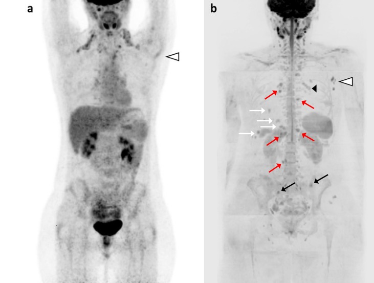
Fig 2. Nodal recurrence or progression to metastatic disease?
36 years old woman with locally advanced ductal breast cancer, after surgery (pT1 N1a M0), local radiation therapy and adjuvant chemotherapy. While under endocrine treatment, an axillary nodal recurrence is diagnosed (histologically proven). In the suspicion of distant metastases, the patient underwent FDG-PET/C. Coronal FDG-PET MIP (a) showed uptake in left axillary lymph nodes (white arrowhead), with no other finding suspicious for metastases. WB-MRI was performed 15 days later: DWI b-900 MIP (b) confirmed the left axillary lymph node metastases (white arrowhead), and detected metastases in bone (left iliac bone, right sacral wing; black arrows), in liver (II and VI segments; white arrows), in lymph nodes (right parasternal, left internal mammary, hepatic hilar, para-aortic and lumbar; red arrows) and in subcutaneous parasternal tissues (black arrowhead). Normal areas of high signal can be seen in cervical and pelvic lymph nodes, brain, spinal cord, spleen, kidneys and in bilateral ovarian cysts. The PD reported at WB-MRI determined a change in treatment from Letrozole to capecitabine, vinorelbine and cyclophosphamide, with additional radiation therapy on bone lesions.

