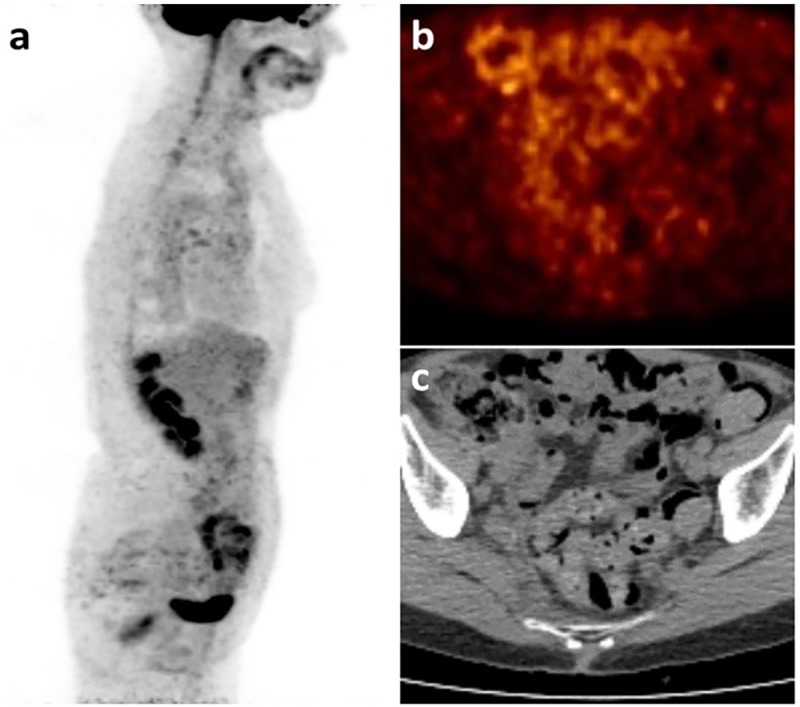
Fig 5. Peritoneal carcinosis undetected at PET/CT.

Patient with locally advanced ductal breast carcinoma (pT2, N2a, M0) after surgery and 4 cycles of chemotherapy, under endocrine therapy. After suspicious rise in CA15-3 the patient underwent FDG-PET/CT, which showed no suspicious uptake and was reported as negative for distant metastases. Sagittal MIP (a) showed non-specific FDG uptake in the ascending colon and tracer excretion in the urinary tract. Pelvic axial cross-section of the FDG-PET/CT (b) and the co-registered CT image (c) did not show any suspicious uptake or measurable lesion.
