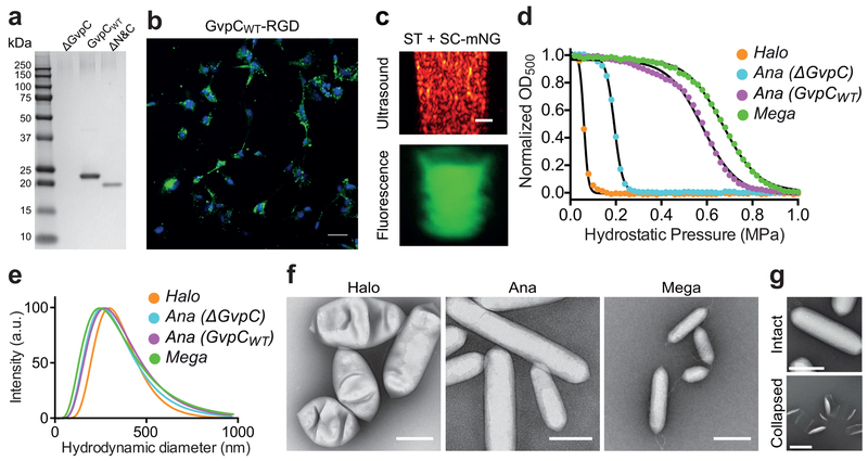
Figure 7. Anticipated results for GV characterization and in vitro ultrasound imaging.
(a) Coomassie stained SDS-PAGE gel indicating complete removal of native GvpC (ΔGvpC, lane 2) and re-addition of two recombinant GvpC variants, GvpCWT and ΔN&C GvpC (lanes 3 and 4). The smeared bands around 250 kD represent GvpA polymers forming the inner GV scaffold whereas GvpC bands are visible at lower molecular weights. (b) Confocal fluorescence image showing dual-functionalized Ana GVs with RGD peptide and Alexa Fluor-488 (green) after a 24 h incubation with U87 glioblastoma cells (DAPI-stained nuclei, blue). Scale bars are 50 μm. (c) Dual-mode imaging of GVs functionalized with SpyCatcher-mNeonGreen with ultrasound (top panel) and fluorescence (ex 506 nm, em 550 nm) (bottom panel). Scale bars are 1 mm. Figures (b) and (c) adapted with permission from Lakshmanan, A. et al. Molecular Engineering of Acoustic Protein Nanostructures, ACS Nano, Copyright © 2016, American Chemical Society. (d) Collapse pressure profiles for GV variants (Halo, Mega, Ana ΔGvpC and Ana GvpCWT). (e) DLS measurements showing typical size distributions for purified Halo, Mega, ΔGvpC and GvpCWT Ana GVs (f) Representative TEM images for purified Halo, Ana and Mega GVs. Scale bars are 200 nm. (g) TEM showing intact and collapsed Ana GVs. Scale bars are 200 nm. Figure (g) adaptedwith permission from Shapiro, M.G. et al. Biogenic gas nanostructures as ultrasonic molecular reporters, Nature Nanotechnology, Copyright © 2014, Nature Research Group.

