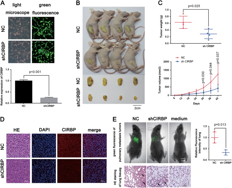
Fig. 3. Knockdown of CIRBP inhibits BCa growth and migration in vivo.
a The green fluorescence of stable cell lines, and qRT-PCR analysis verification of CIRBP knockdown efficiency by lentiviral-CIRBP-shRNA. b Xenograft models (n = 4) were established by subcutaneously inoculating LV-NC cells or LV-shCIRBP cells and allowed to grow for 6 weeks, then the mice were sacrificed and the tumors were taken out and weighed. c tumor volume measurement and tumor weight. d Representative H&E staining and immunefluorescence staining of xenograft tumors from the tumor-bearing mice of the LV-NC group and LV-shCIRBP group, indicating the downregulation of CIRBP (red) in LV-shCIRBP group. Nuclei were stained by DAPI (blue). e Pulmonary metastasis models (n = 3) were established by tail intravenous injecting LV-shCIRBP UM-UC-3 cells or LV-NC UM-UC-3 cells, the fluorescence intensity of pulmonary metastasis tumor was measured to evaluate the migration capacity. Representative H&E staining of lung tissues indicating the pulmonary metastasis tumors (pointed by the arrows). Statistical analysis of the fluorescence intensity was calculated using T-test. Means ± standard deviation from three independent experiments

