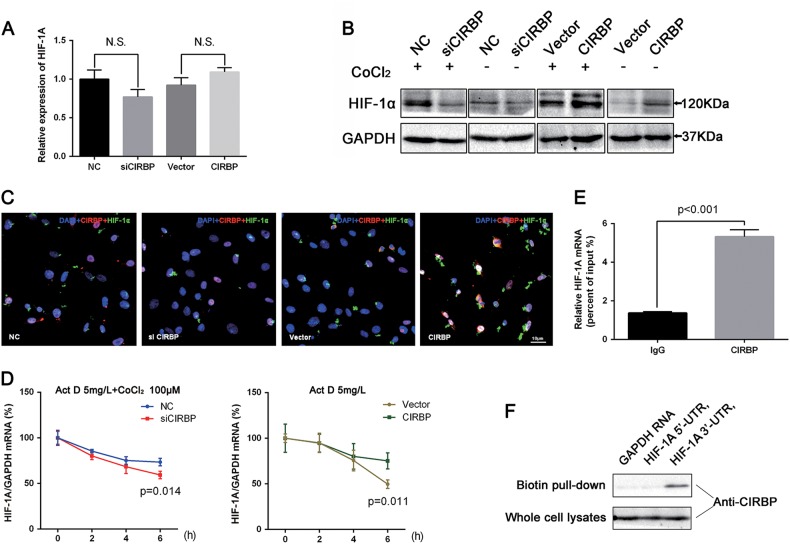
Fig. 4. CIRBP increases mRNA stability and protein translation of HIF-1α in BCa cells.
a qRT-PCR analysis for the effects on the HIF-1A mRNA levels of CIRBP knockdown and CIRBP overexpression. b Western blot analysis of HIF-1α performed on CIRBP knockdown and CIRBP overexpression UM-UC-3 cells, exposed (+) or not (−) to CoCl2 (100 μM for 4 h, Sigma 15862). c Immunofluorescence staining revealed alterations of HIF-1α (green) after 48 h transfection of siCIRBP in UM-UC-3 cells or CIRBP overexpression plasmid in UM-UC-3 cells under normoxia (CIRBP red). Nuclei were stained by DAPI (blue). d 24 h after transfection with siCIRBP (pretreated with CoCl2) or CIRBP overexpression plasmid (under normoxia), UM-UC-3 cells were cultured in the presence of Actinomycin D (15 μg/ml, Abcam ab141058), qRT-PCR detected mRNA levels of HIF-1A specific time points of 0, 2, 4, 6 h. GAPDH is used as the normalization control. e RNA-Binding Protein Immunoprecipitation (RIP) assay was performed with anti-CIRBP or anti-IgG, qRT-PCR detected the IP efficiency (percent input). f Biotin pull-down assay was achieved to confirm the interacts of CIRBP and HIF-1A mRNAs 3′-UTR, both 3′-UTR and 5′-UTR of HIF-1A mRNA transcripts were constructed with biotin-label, transcripts of GAPDH mRNA were used as a negative control. Means ± standard deviation from three independent experiments. T-test was used for statistical analysis

