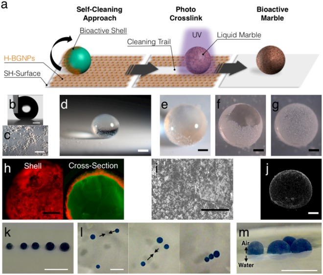
Figure 2.
Fabrication of the hydrogel marble. (a) Schematic drawing of the synthesis procedure of the bioactive hydrogel marble by a biomimetic approach based on the self-cleaning of the lotus leaf. (b) Profile of a water drop in contact with the superhydrophobic surface. Water contact angle of 162 ± 1.3°. Scale bar = 500 µm. (c) Superhydrophobic surface covered with H-BGNPs. The observed clusters could be attributed to powder electrostatic effects. Scale bar = 1 cm. (d) Polymeric drop on the superhydrophobic surface. Scale bar = 500 µm. (e) Detail of the polymeric drop rolling on the superhydrophobic surface. Scale bar = 500 µm. (f) Formation of the bioactive shell. Scale bar = 500 µm. (g) Bioactive marble: polymeric sphere covered with H-BGNPs. Scale bar = 500 µm. (h) Fluorescent micrographs detail of the shell and cross-section of the bioactive marble. Before the fabrication of the bioactive marble, the gelatin and the H-BGNPs were first stained with fluorescein (green) and rhodamine (red), respectively. Scale bar = 500 µm. (i) Representative SEM micrographs of the H-BGNPs at the surface of the BHM. Scale Bar = 5 µm. (i) μCT 3D reconstruction images of the BHM. (k) Photographs showing BHM (dyed in blue) on top of a superhydrophobic surface. The dispensed volumes were 1.5, 2, 4, 8, and 16 μL. The approximate diameters of the spheres after crosslinking were 1, 1.2, 2.5, 4; 5; 6 mm, respectively. Scale bar = 1 cm. (l) BHM floating at the surface of the water. Scale bar = 0.5 cm. (m) Representative image of the floating performance of the BHM cluster that could be manipulated and self-assemble on the surface of the water. Scale bar = 1 cm.

