Figure 2.
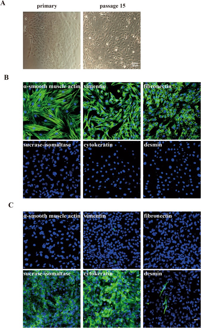
Morphological characterization of the myofibroblasts isolated from porcine ileum. (A) Characteristics of porcine ileum myofibroblasts. Myofibroblasts could be observed at 5 days after isolation. They had a cobblestone like morphology. They could be further sub- passaged as a continuous cell line. (B) A third passage of myofibroblasts was identified by immunofluorescence staining. Cells stained characteristically for intestinal myofibroblasts. They were positive for α-smooth muscle actin, vimentin and fibronectin, while cells were negative for sucrase-isomaltase, cytokeratin and desmin. (C) Primary ileum epithelial cells were used as control and stained with all markers that were used for the staining of the myofibroblasts. Scale bar: 25 µm.

