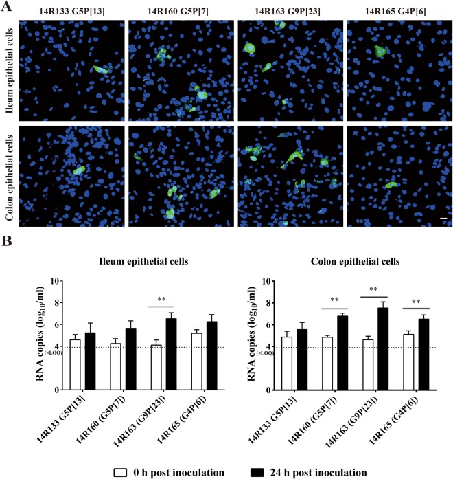
Figure 7.
Susceptibility of porcine primary enterocytes to different rotavirus genotypes present in fecal suspension of diarrheic pigs. The infected cells were visualized by immunofluorescence staining at 24 h post inoculation (A). Viral RNA titer with supernatant of co-cultured primary porcine enterocytes inoculated with different rotavirus fecal suspensions at 0 h and 24 h post inoculation (B). Scale bar: 25 µm. Data are expressed as mean ± standard deviation of the results of 3 separate experiments. Statistically significant differences in comparison with the data from 0 h p.i. are represented as *P <0.05 or **P <0.01. <LOQ: below limit of quantification.

