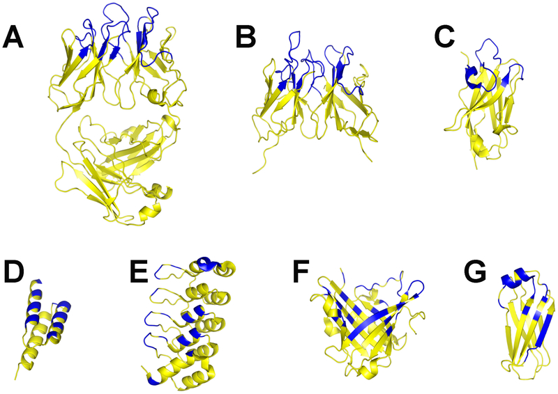
Figure 3. Chaperone platforms for membrane protein co-crystallization (yellow), depicting relative sizes and binding surfaces (blue).
(a) Fab fragment (PDB code 3EFF) [50]. (b) scFv (PDB code 3NN8) [78]. (c) Camelid nanobody (PDB code 2P42) [73]. (d) Affibody (PDB code 3MZW) [74]. (e) DARPin (PDB code 2J8S) [51]. (f) Anticalin (PDB code 3BX7) [76]. (g) FN3 domain (PDB code 2OCF).

