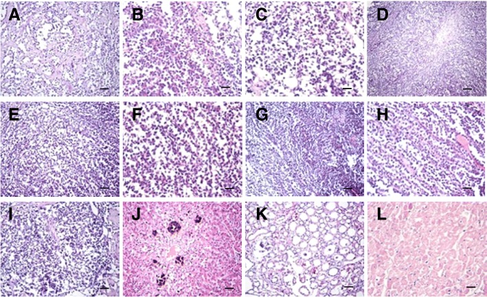
Fig. 4.
Histopathological results of main lesion. a-c Severe alveolar atrophy or even disappearance, pulmonary fibrosis hyperplasia in the lung, visible tumor cell metastasis. d-f Ovarian tissue showed a large number of round cells were nests or cord-like proliferation, spindle-shaped cells proliferation in the periphery, accompanied by the formation of collagen fibers, tumor cell atypia is not obvious, rare cells split phase, local necrotic calcification area. g-h Perianal tissue showed tumor cell metastasis accompanied by fibrogenesis. i Fewer spleen lymphocytes than normal, monocyte proliferation. j Severe hepatic steatosis, partial necrosis, multifocal calcification, small focal inflammatory infiltration of the liver. k Part of glomerular atrophy, mild glomerular swelling, tubular significant dilatation, visible proteinuria and cell tube in the kidney. l Severe swelling of myocardial cells, granular degeneration with partial necrosis in the heart. (d: Bar = 100 μm. a, e, g, i, j and k: Bar = 50 μm. b, c, f, h and l: Bar = 20 μm)

