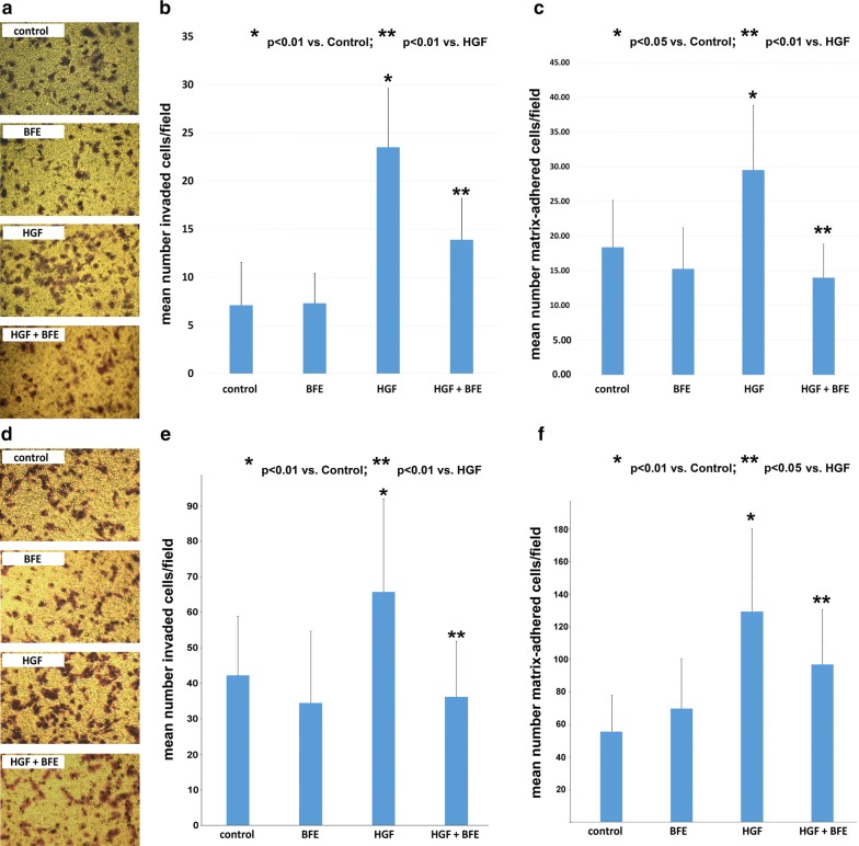
Fig. 2.
HGF-stimulated breast cancer cell invasion and cell–matrix adhesion was inhibited through BFE treatment: a BT549 cell line; d MDA-MB-231 cell line—Images taken following a 48 h incubation period using the breast cancer cell invasion model. The enhanced degree of invasion through HGF stimulation was observed through the higher level of successfully invaded breast cancer cells when compared to the control group. BFE was able to suppress this effect when incubated with HGF, and importantly addition of BFE alone had no bearing on the degree of breast cancer cell invasion. b BT549 cell line; e MDA-MB-231 cell line—the number of cells that successfully invaded were quantified and results plotted to reveal the statistically significant increase in cell invasion following HGF treatment, which was significantly inhibited through BFE treatment of breast cancer cells. c BT549 cell line; f MDA-MB-231 cell line—the number of breast cancer cells that formed a strong attachment to a replica matrix during the allotted time was quantified and the data reveals that HGF significantly enhanced cell–matrix adhesion and that the edition of BFE was able to quench the degree of attachment by these cells

