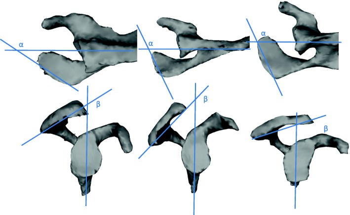
Fig. 2.
3-D reconstruction of the segmented scapulae showing distinct differences regarding relative external acromial rotation (α – angle between a tangent to the lateral acromial border and a line parallel to the scapular body) and posterior acromial slope (β- angle between a line connecting the posteroinferior and the anteroinferior acromion and a line parallel to the scapular body). From left to right “p1” scapula, “p2” scapula, “p3” scapula

