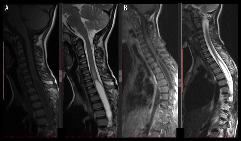
Figure 5.
Postoperative 3- and 6-month MRIs of the same patient as in Figure 4. She underwent NTR of the lesion using T8-10 laminoplasty. No adjuvant treatment was carried out for the syrinx. Both MRIs showed regress of syrinx cavities, and no progression or seeding metastasis was detected; (A) Postoperative 3-month MRI, T1-weighted MRI in left side and T2-weighted MRI in right side; (B) Postoperative 6-month MRI, T1-weighted MRI in left side and T2-weighted MRI in right side. Note that the syrinx cavities were regressed without additional therapy.

