
Figure 4. pUR4 decreases proliferation and migration of human failing cardiac fibroblasts.
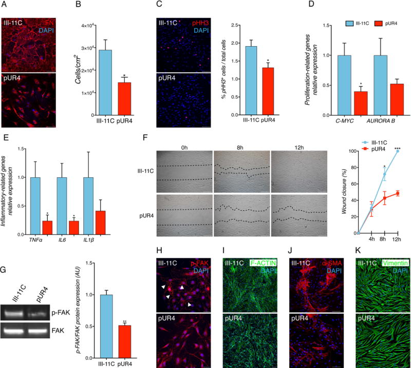
(A) pUR4 reduces FN staining. Scale bars, 100 μm. (B) pUR4 decreases cell proliferation. (C) pUR4 decreases phospho- histone H3 (pHH3)-positive human heart failure CF (representative pictures, left, and quantification, right). Scale bars, 100 μm. (D) pUR4 downregulates proliferation-related genes. (E) Expression levels of pro-inflammatory cytokine transcripts are significantly reduced following pUR4 treatment in failing human cardiac fibroblasts measured by RT-qPCR. Tumor necrosis factor alpha (TNFα), Interleukin 6 (Il6) and Interleukin 1ß (IL1ß). (F) Cardiac fibroblasts were scratch-wounded and recorded at 0 h, 4 h, 8 h and 12 h post-scratch. Black dotted lines denote the wound borders (representative photographs, left, and mobility quantification, right). Scale bars, 500 μm. (G) p-FAK protein level expression (representative immunoblots, left, and densitometry, right). (H) Immunofluorescence analysis of p-FAK in pUR4 and III-11C-treated human MF (arrowheads). Scale bars, 10μm. (I) F-actin staining in pUR4-treated cells indicated a compromised defect in cellular organization compared to control group. Scale bars, 100 μm. (J) Alpha-smooth muscle actin (α-SMA) staining in III-11C and pUR4-treated human MF. Scale bars, 100 μm. (K) Vimentin staining in peptide-treated cells. Scale bars, 100 μm. Statistical significance was determined with paired t-test: *P<0.05, **P<0.01, ***P<0.001. Data are represented as mean ± SEM. n=5.