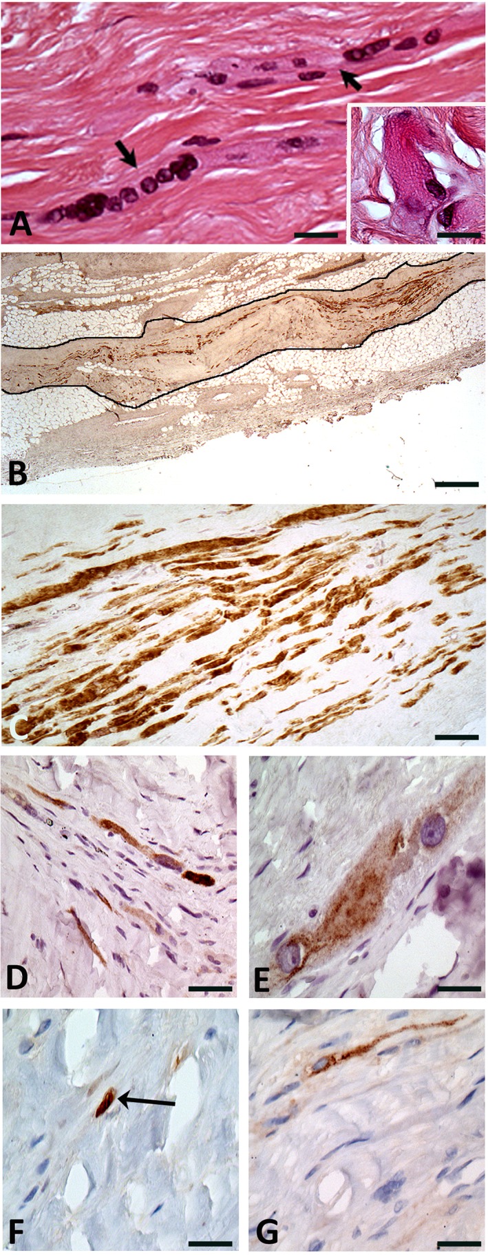
Figure 1.

Histological and immunochemical findings in the native heart at the site of the cell‐transplanted myocardial infarction. (A): Multinucleated skeletal muscle cells/myotubes (arrows) embedded in fibrosis. Inset shows the Z‐bands in a skeletal muscle cell. Hematoxylin & Eosin staining. Original ×40 and ×100 for inset. (B): Large sub‐epicardial focus (black outline) of grafted skeletal muscle cells expressing the fast isoform of the skeletal muscle myosin heavy chain. Original ×2.5. (C): Strong expression of the fast isoform of the skeletal muscle myosin heavy chain (MYH1) by several cells. Although the density of skeletal muscle cells is high, they are separated by fibrosis. Original ×20. (D): Expression of the slow isoform of the skeletal muscle myosin heavy chain (MYH7) by a small proportion of the grafted cells. Original ×20. (E): Expression of the skeletal troponin T (TNNT3) fast isoform by the grafted skeletal muscle cells. Original ×40. (F): Expression of the myogenin transcription factor in nuclei of the grafted skeletal muscle cells (arrow). Original ×40. (G): Expression of CD56 by myotubes. Original ×40.
