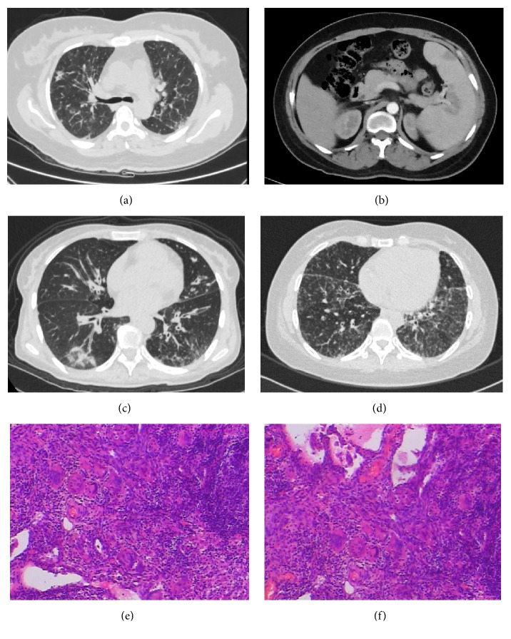
Figure 1.
CT appearance of the three cases and pathological features of case 3. Panel (a) showed diffused reticulation as the manifestation of suspected GLILD in case 1. Panel (b) showed focal hypodense splenic lesions with splenomegaly in case 1. Panel (c) showed bronchiectasis with infiltrates and mucus plugs in case 2. Panel (d) showed diffused micronodules and bronchiectasis as the manifestation of GLILD in case 3. Panels (e) and (f) revealed diffused infiltration of lymphocytes and lymphoid follicles formation in the lung tissues, scattered with epithelioid granulomas and multinuclear giant cells, consistent with the pathological manifestation of GLILD.

