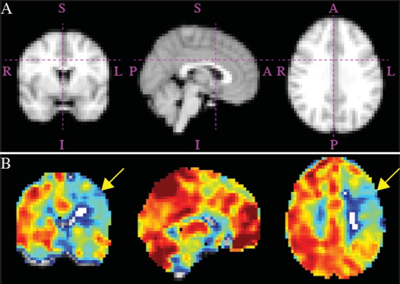
Figure 1.

Admission MRI studies obtained in a patient with a symptomatic left hemisphere. Corresponding atlas maps for hemodynamic sections (a) and orthogonal representations of reactivity maps (b), demonstrating impairment in CVR in the left hemisphere (yellow arrows). Right hemisphere (asymptomatic) PIRAMD score: 0 (Grade 1); left hemisphere (symptomatic) PIRAMD score: 10 (Grade 3). A = anterior; I = inferior; L = left; P = posterior; R = right; S = superior. Figure is available in color online only.
