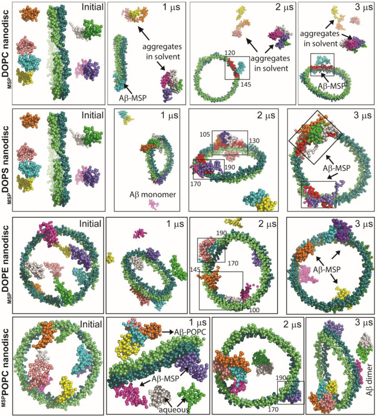
Fig. 5.
MD simulations reveal Aβ40-MSP interactions. Illustration of Aβ40 interaction with MSP in nanodiscs at the indicated MD simulation times. MD snapshots are retrieved after every 1 μs from zwitterionic (DOPC/DOPE/POPC) or anionic (DOPS) MSP-based lipid-nanodiscs. The MSP protein chains (ring shape) and Aβ molecules (eight) are represented in vdw using VMD and drawn in different colors. The lipid molecules in the center and water inside the box are not shown for visual clarity. MSP domains binding with Aβ40 are covered in a rectangle and the amino acid residues spanning the binding domains are indicated. All Aβ40 molecules are initially placed ≈ 10 Å away from the lipid bilayer surface and a minimum distance of « 5 Å is maintained between Aβ molecules in aqueous phase.

