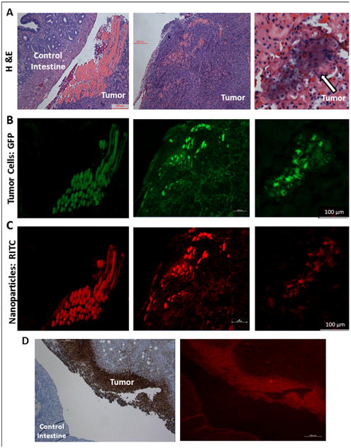
Figure 8.

A. Mouse tumor tissue sections stained with hematoxylin and eosin (H&E) stain after necropsy reveal significant tumor burden. B. GFP fluorescence reveals tumor cell location. C. RITC fluorescence reveals highly specific nanoparticle tumor targeting; MSN-HA nanoparticles are only localized in tumor cells and not in control intestine. D. MSN-HA co-localizes with CD44 on the membrane of tumor.
