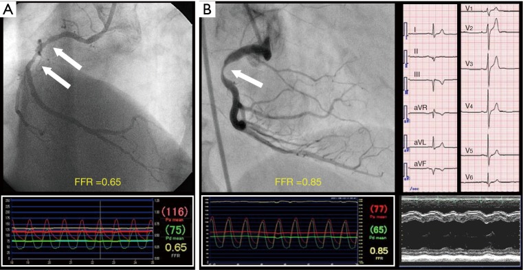
Figure 2.
Mismatch between angiographic DS and FFR. (A) Positive mismatch (FFR ≤0.80 and DS <50%); (B) negative mismatch (FFR >0.80 and DS ≥50%) due to small amount of viable myocardium. Electrocardiogram shows QS pattern in II, III, aVF leads. Echocardiogram shows akinesis in left ventricular inferior wall. Arrow indicates a stenosis. DS, diameter stenosis; FFR, fractional flow reserve.

