
FIGURE 5.
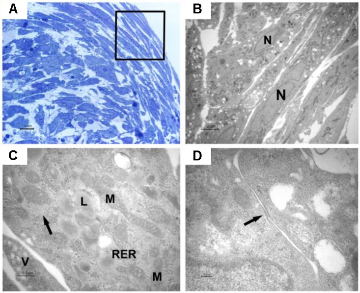
Light and transmission electron microscopy of EB-hGMSCs periphery. (A) Light microscopy of semithin section stained with toluidine blue at high magnification. The square marked peripheric area where the cells are elongated. Bar: 10 μm. (B–D) Transmission electron microscopy. (B) A magnification of an area similar to that marked by square in (A) shows some layers of elongated cells with euchromatic nuclei (N). Bar: 2 μm. (C) At higher magnification, filaments (arrow), lipid droplets (L), mitochondria (M), rough endoplasmic reticulum (RER), and vesicles (V) are visible in the cytoplasm. Bar: 0.2 μm. (D) Plasma membrane thickening is present (arrow). Bar: 0.1 μm.