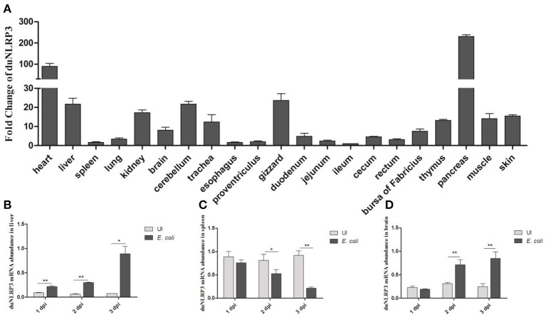
Figure 2.
Expression profiles of NLRP3 in Cherry Valley duck. (A) Expression of duNLRP3 in tissues of healthy Cherry Valley duck, the ileum was chosen as the standard tissue. Expression of duNLRP3 in the (B) liver, (C) spleen, and (D) brain of Cherry Valley ducks from E. coli uninfected group (UI) and E. coli infected group (E. coli), and these mRNA fold changes were calculated using the E. coli-infected ducks vs. control group ducks at the same time point. The relative expressions of duNLRP3 were normalized by β-actin with the 2−ΔΔCt (A) and 2−ΔCt (B–D) method. Means ± SD (n = 3) from three independent repetitions are presented. The student's t test was performed to evaluate the differences. *Significant difference (P < 0.05); **highly significant difference (P < 0.01); dpi, days post-infection.

