
Figure 5. Heat shock protein (Hsp) 70.
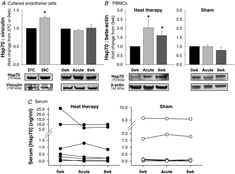
A and B, Hsp70 in (A) cultured endothelial cells pre‐incubated at 37°C, at 39°C (heat pretreatment) or with serum from human subjects collected before (0wk) and after (Acute HT) the first hot water immersion session or following 8 weeks of heat therapy (8wk) and in (B) peripheral blood mononuclear cells collected at the same time points from human subjects who participated in 8 weeks of heat therapy (left) or 8 weeks of thermoneutral water immersion (right). Representative Western blot images are shown below. Data are mean ± SEM. C, free Hsp70 concentrations in serum collected from human subjects at the same time points (n = 6‐7 per group; individual data shown). [Hsp70] was below detection in several subjects (excluded from analysis; n = 3 in heat therapy group, n = 4 in sham group). Despite high variability across individuals, [Hsp70] concentrations were relatively stable within individuals across the interventions.