Fig. 2.
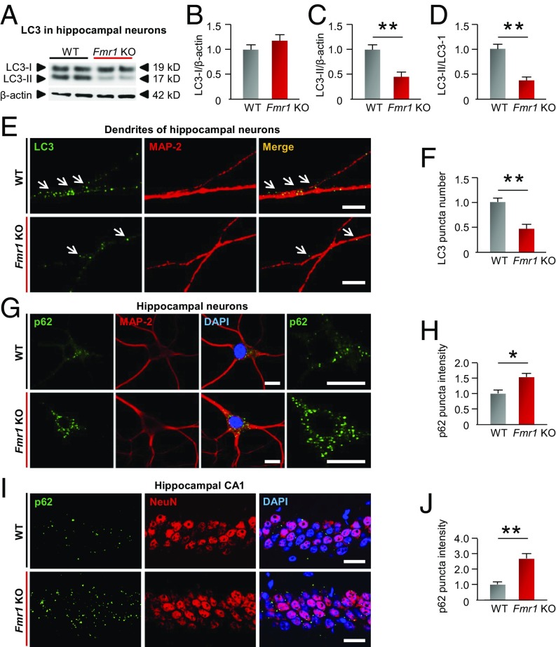
Autophagy is down-regulated in hippocampal neurons of fragile X mice. (A) Representative Western blots of whole-cell lysates from primary cultures of hippocampal neurons at DIV 14 from WT and Fmr1-KO mice were probed for LC3-I and LC3-II. β-Actin was used as a loading control. (B–D) Summary data showing LC3-I (B), LC3-II (C), and the LC3-II/I ratio (D) (normalized to the corresponding WT values). (E) Green immunofluorescence (LC3+ puncta) marks autophagosomes (arrows) localized to MAP-2–labeled dendrites (red) in neurons from KO and WT mice. (Scale bars, 10 µm.) (F) Summary data showing number of green (LC3+) puncta colocalized with red (MAP2+) in dendrites (normalized to WT group). (G) Images show immunolabeling of p62 together with MAP-2 to mark neurons. p62 staining at higher magnification is shown in Right panels. (Scale bars, 15 µm.) (H) Summary data (bar graphs) showing fluorescence intensity (normalized to WT values). (I) Five-week-old male WT and Fmr1-KO mice were killed, and frozen sections of brain were subjected to immunolabeling with an antibody to p62 together with an antibody to NeuN to mark neurons. (Scale bars, 25 µm.) (J) Summary data show the calculated intensity (normalized to corresponding WT values). *P < 0.05, **P < 0.01. Significance was calculated by a two-tailed Student’s t test; n = 5 experiments involving independent batches of neurons cultured from different litters in B–D, F, and H. At least 50 neurons were analyzed for each treatment group in F and H. n = 4 mice for J. Values reflect mean ± SEM.

