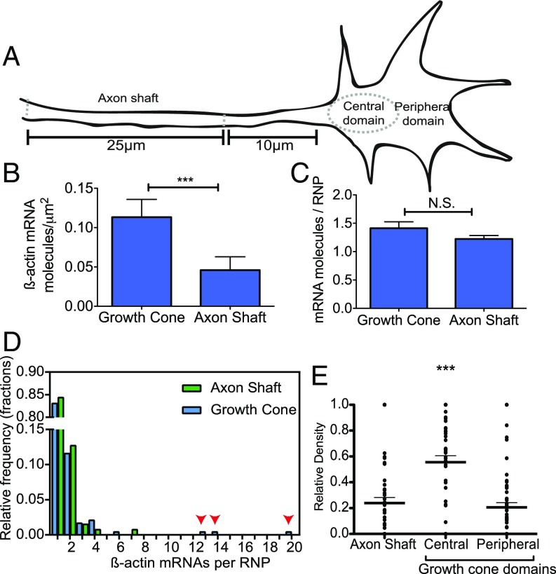
Fig. 4.
smFISH reveals that the localization patterns of β-actin mRNA vary according to axonal subcompartment. (A) Cartoon representation of the different axonal regions analyzed. (B) A comparison of the density of β-actin mRNA molecules in the growth cone and axon shaft via smFISH shows significantly increased density of β-actin mRNA in the growth cone. (C) No difference between β-actin mRNA RNP stoichiometry was observed in the different subcompartments. N.S. not significant. (D) Histogram of β-actin mRNA stoichiometry. Arrowheads indicate more highly multiplexed copy numbers in the growth cone. (E) Relative density of β-actin mRNA across subcompartments of the same axon shows significant enrichment in the central domain of the growth cone. ***P < 0.0001; paired Student’s t test; n = 63 axons.

