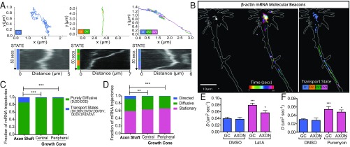
Fig. 5.
Differential subcompartmental trafficking of β-actin mRNA. (A) Examples of complex motion trajectories displayed by β-actin mRNA in axons. Different transport modes were characterized through HMM–Bayes. D, diffusive states; DV, directed transport states. (B) β-Actin mRNA dynamics within a representative axon. (Left) Initial β-actin mRNA distribution labeled by MBs. (Center) Temporally color-coded β-actin mRNA dynamics. (Right) Annotated mRNA trajectories. (C) Significantly more trajectories undergo motion with directed transport in the axon shaft than in the central and peripheral domains of the growth cone. (D) Trajectories annotated according to the predominant motion state again show a significantly greater fraction of directed transport of β-actin mRNA in the axon shaft. ***P < 0.001, **P < 0.0025; χ2 test; n = 6, 66 axons. (E) The addition of latrunculin A significantly increased the diffusive mobility of MB β-actin mRNA puncta compared with DMSO treatment only [***P < 0.0001 and *P = 0.0454 for growth cone (GC) and axon shaft; n = 12 axons for DMSO treatment, and n =11 axons for latrunculin A]. (F) Dissociation from ribosomes by puromycin addition increased the diffusion coefficient of MB puncta in both axons and growth cones compared with wild-type untreated axons [n = 11 puromycin-treated axons and n = 66 wild-type axons, ***P < 0.0001 (growth cone) and *P = 0.0253 (axon)].

