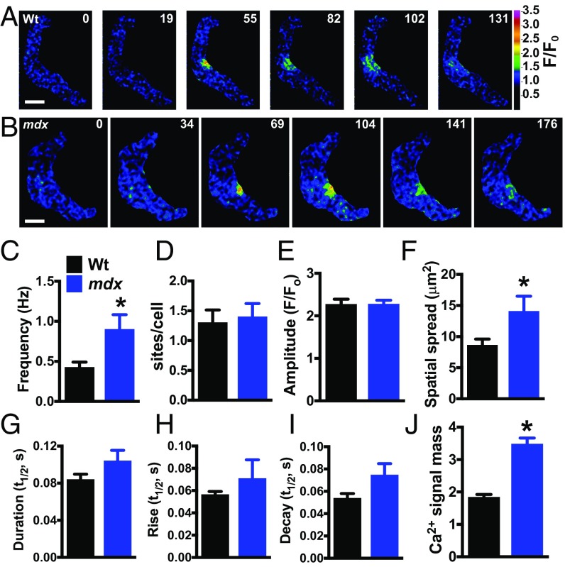
Fig. 3.
Spontaneous Ca2+ spark frequency and signal mass are elevated in SMCs from mdx mice. (A and B) Pseudocolored confocal Ca2+ images of SMCs from wild-type (A) and mdx (B) mice showing representative time courses of the fractional increase in fluorescence (F/F0) during a typical Ca2+ spark event. (Scale bars, 10 µm.) (C–I) Summary data showing frequency (Hz), number of Ca2+ spark sites per cell, amplitude (F/F0), area of spatial spread (µm2), half-duration [half-time (t1/2), s], rise time (t1/2, s), and decay time (t1/2, s) of Ca2+ sparks in SMCs from wild-type and mdx mice. The frequency and area of spatial spread of Ca2+ sparks in SMCs from mdx mice were greater than those from wild-type mice (n = 9 cells per group, five animals per group; *P < 0.05). (J) Ca2+ signal mass for each Ca2+ spark event was greater in SMCs from mdx mice compared with wild-type (n = 170 to 285; *P < 0.05). All data are mean ± SEM.

