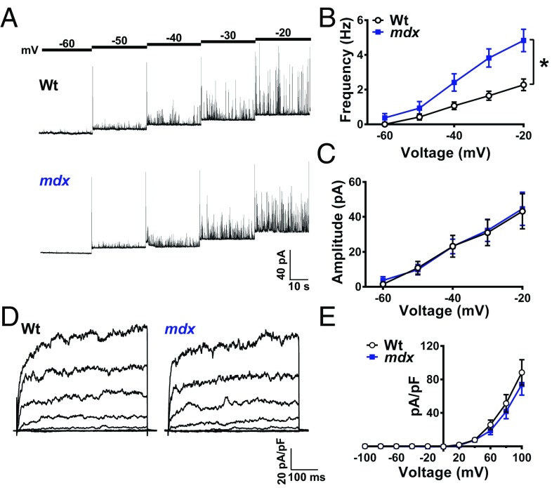
Fig. 4.
Spontaneous Ca2+-activated K+ channel activity is elevated in SMCs from mdx mice. (A) Representative traces of STOCs recorded from SMCs from wild-type and mdx mice over a range of membrane potentials (−60 to −20 mV). (B) Summary data showing that STOC frequency is greater in SMCs from mdx mice (n = 14 cells per group, five animals per group; *P < 0.05). (C) STOC amplitude did not differ between groups (n = 14 cells per group, five animals per group. (D) Representative conventional whole-cell patch-clamp recordings of paxilline-sensitive BK currents in SMCs from wild-type and mdx mice. Currents were recorded during a series of command voltage steps (−100 to +100 mV). (E) Summary of whole-cell current data (n = 7 cells per group, three animals per group). There were no significant differences. All data are mean ± SEM.

