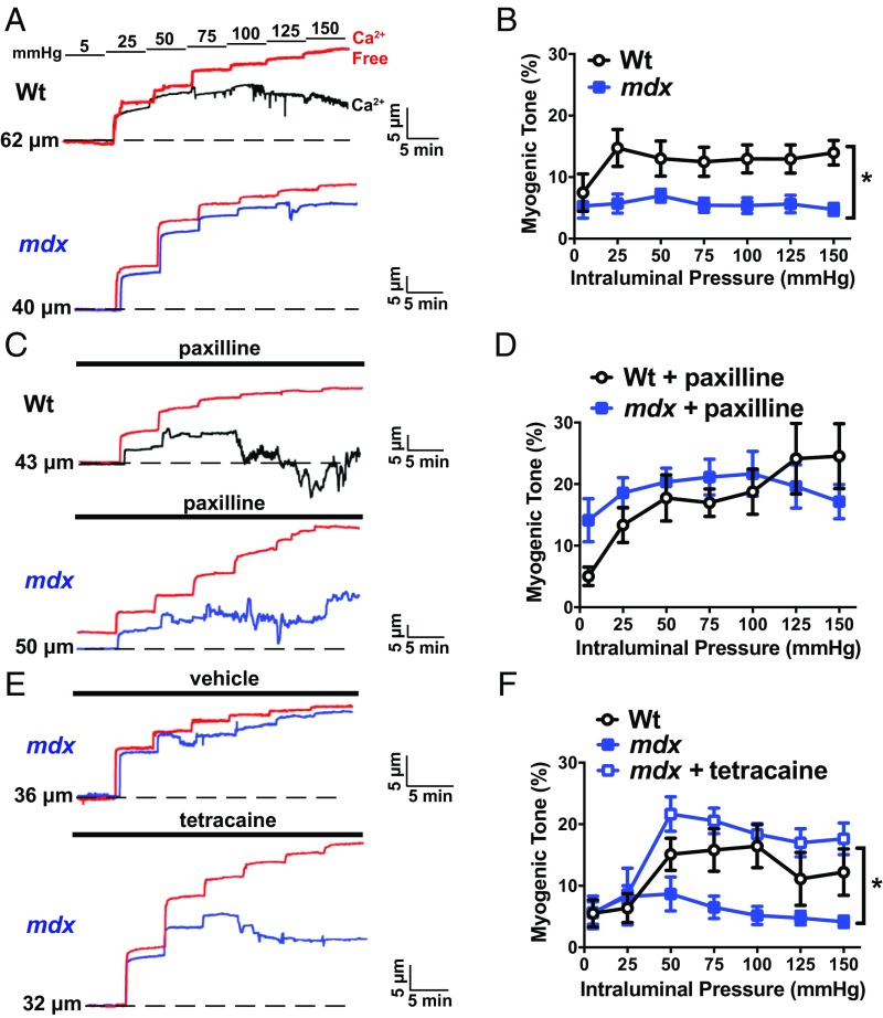
Fig. 5.
Functional impairment in cerebral pial arteries from mdx mice. (A) Representative traces showing changes in luminal diameter over a range of intraluminal pressures (5 to 150 mmHg). (A, Top) A recording obtained from a cerebral pial artery from a wild-type mouse (black). (A, Bottom) A recording from an artery from an mdx mouse (blue). In both panels, the passive change in diameter in response to intraluminal pressure is shown in red. (B) Myogenic tone for both groups (n = 7 or 8 arteries per group, four animals per group; *P < 0.05). Myogenic tone was greater in cerebral arteries from wild-type mice than those from mdx mice. (C) Similar experiment to that shown in A, but in the presence of the BK channel blocker paxilline (1 µM). (D) Mean myogenic tone in the presence of paxilline (n = 7 or 8 arteries per group, four animals per group). There were no significant differences. (E) Representative traces showing the effects of blocking ryanodine receptors with tetracaine (10 µM) on the myogenic tone of cerebral pial arteries from mdx mice. (F) Mean myogenic tone of arteries from wild-type mice and mdx mice in the presence and absence of tetracaine (n = 7 arteries per group, four animals per group; *P < 0.05). All data are mean ± SEM.

