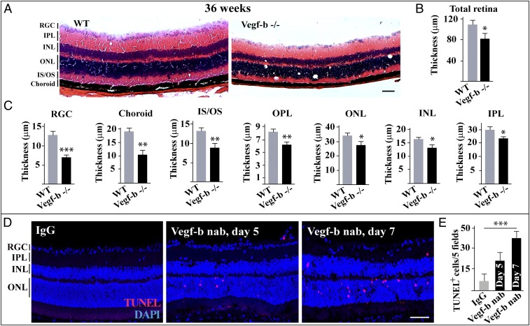
Fig. 1.
Genetic deletion of Vegf-b leads to retinal degeneration. (A–C) H&E staining shows that the thickness of the retinal layers of 36-wk-old Vegf-b−/− mice was significantly reduced, including the retinal ganglion cell layer (RGC), choroid, inner segment/outer segment (IS/OS), outer plexiform layer (OPL), outer nuclear layer (ONL), inner nuclear layer (INL), and inner plexiform layer (IPL) (n = 8; ***P < 0.001, **P < 0.01, *P < 0.05). (D and E) TUNEL staining shows that intravitreal injection of Vegf-b nAb into the vitreous of normal mice led to retinal apoptosis 1 wk after injection (n = 8; ***P < 0.001). (Scale bar: 50 µm.)

