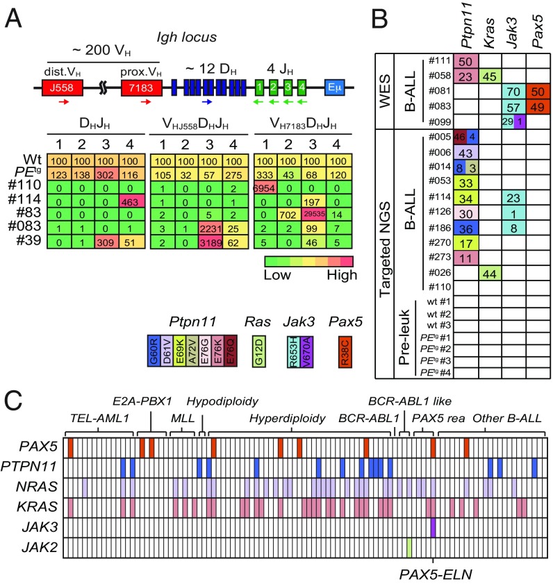
Fig. 2.
Clonal selection in PAX5-ELN–induced B-ALL. (A) Schematic of the mouse IgH locus (Upper). The locus is composed of ∼200 VH (red boxes), ∼12 DH (blue), and 4 JH (green) segments that undergo recombination in B-cell precursors to produce a functional VHDHJH unit. Genomic DNAs were prepared from purified PEtg pro-B cells and five leukemic PEtg mice and subjected to quantitative PCR to quantify DHJH and VHDHJH rearrangements using primers that bind the indicated gene segments. dVH, distal VH; pVH, proximal VH. PCR of the HS5 element downstream of the 3′ regulatory region was performed for normalization of DNA input. PCR was performed in triplicate. Quantification of DHJH and VHDHJH rearrangements is represented as a heat map (Lower). DNA from purified WT pro-B cells was used as a control and normalization (100% of the signal) for each gene rearrangement ranked in each column. (B) Recurrent mutations in B-ALL blasts induced by PAX5-ELN. Whole-exome sequencing (WES) was performed on BM cells from five leukemic PEtg mice. Mutations found on Ptpn11, Kras, Pax5, and Jak3 genes were verified and screened for recurrence by targeted next-generation sequencing (NGS) on BM cells from 11 leukemic, 3 WT, and 4 preleukemic 30-d-old PEtg mice. Each row represents a leukemia sample, and each column represents a genetic alteration. Colors indicate the position of the mutation, and numbers represent the variant allele frequency of each mutation. (C) Mutations on PTPN11, KRAS, NRAS, JAK3, JAK2, and PAX5 genes were screened for recurrence on 101 B-ALL patient samples. Each column represents a B-ALL sample classified according to the indicated oncogenic subtypes, and each row represents a genetic alteration. Colored boxes indicate the presence of a mutation, listed in Dataset S1.

