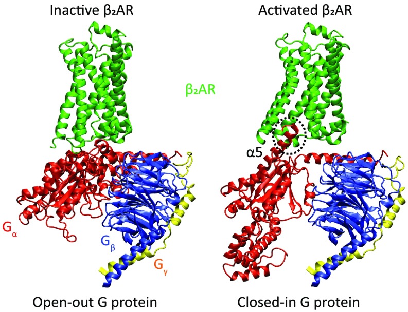
Fig. 2.
Protein structures of representative assemblies (the selection criterion was visual clarity) used in this study (see main text and Materials and Methods for details). The receptor is shown in green, and the G protein α-, β-, and γ-subunits are shown in red, blue, and yellow, respectively. The G-protein α-subunit α5 helix mentioned in the text is labeled and its C terminus, which undergoes a disorder-to-order transition, is marked with a dashed circle (where it is ordered, or “in”). Note that the specific complex in PDB ID code 3SN6 is the activated receptor bound to open-in G protein, which is not shown here.

