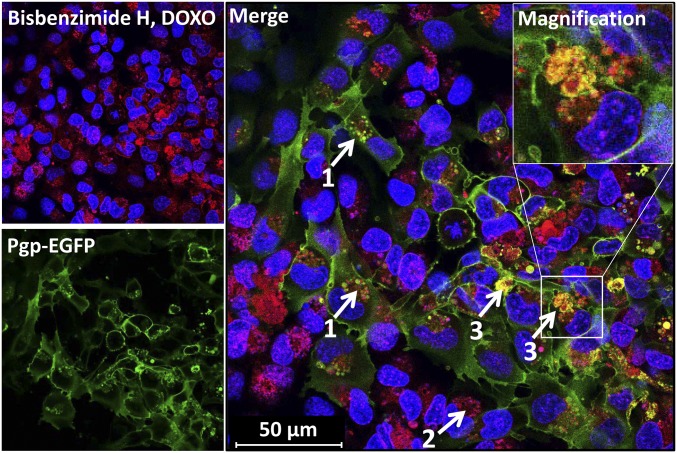
Fig. 2.
Intracellular Pgp/Pgp substrate vesicle and barrier-body formation after exposure of BCECs to DOXO. Cocultured hCMEC/D3-MDR1-EGFP and hCMEC/D3 WT cells were treated with DOXO (10 µM, 30 min) and subsequently analyzed by live cell imaging and confocal microscopy. DOXO (red) is enriched in Pgp-EGFP positive (green) intracellular vesicles of EGFP-overexpressing cells (1). Likewise, DOXO accumulates in vesicular structures near to cell nuclei of WT cells (no green fluorescence) (2). Like the results from EFIG-AM treatment of hCMEC/D3 cells, accumulation of Pgp/DOXO-enriched vesicles (barrier bodies) can be observed at the plasma membrane borders of the cells (3). (Inset) Magnification of barrier body (white frame, merged image). For orientation, cell nuclei were stained by the DNA intercalating dye bisbenzimide H (blue).

