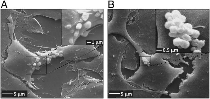
Fig. 4.
Vesicle formation and aggregation at the apical surface of human BCECs after treatment with DOXO. hCMEC/D3 cocultures were grown on collagen-coated coverslips in 24-well cell culture plates. After treatment with DOXO (10 µM, 30 min), cocultures were fixed with 2.5% glutaraldehyde for analysis by scanning electron microscopy. (A) A scanning electron micrograph showing vesicle formation at the apical plasma membrane of hCMEC/D3 cells after treatment with DOXO. Vesicle size in diameter: 1–2 µm. [Magnification: 2,000× (main image) and 10,000× (Inset).] (B) Representative scanning electron micrograph showing aggregation of EVs at the apical surface of hCMEC/D3 cells, thus forming aciniform barrier bodies. The single vesicle size in diameter ranged from 0.6 to 2 µm, and the size of EV aggregates in diameter ranged from 5 to 16 µm. [Magnification: 2,000× (main image) and 20,000× (Inset).]

