Fig. 5.
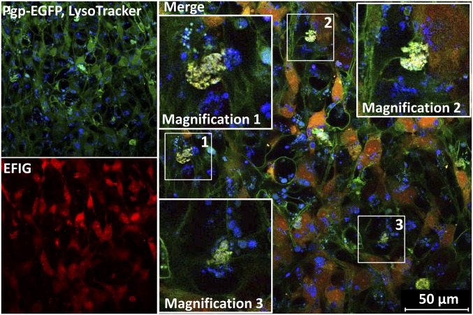
Barrier bodies show lysosomal staining. Lysosomes of hCMEC/D3 cocultures were stained by incubation with 75 nM LysoTracker (blue) before EFIG-AM treatment (30 min) and subsequently analyzed via live cell imaging and confocal microscopy. Formed barrier-body aggregates, yellow in merged images (Pgp-EGFP, green; EFIG, red), additionally exhibit a blue fluorescent signal, suggesting a lysosomal origin of these structures. (Insets) Magnification (2.5-fold) of barrier bodies 1–3 (white frames in merged image).

