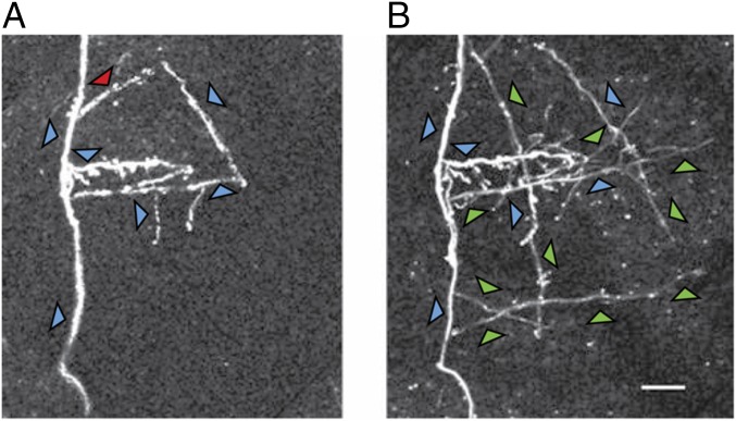
Fig. 3.
Two-photon image of a selected region of cortex taken at the beginning of training (A) and the same region imaged 8 wk later during the course of training (B). Between the two time points, several axon collaterals have sprouted. Axons that were stable over the time period are indicated by blue arrows, a pruned axon collateral is indicated by the red arrow, and newly sprouted axons are indicated by the green arrows.

