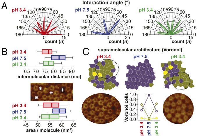
Fig. 2.
Analysis of the supramolecular structure of GLIC channels. (A) Polar coordinates plot of the intermolecular connections in Fig. 1F. At pH 7.5, the molecules engage in an ordered hexagonal packing with only three angles with 60° difference populated. At pH 3.4 (beginning and end of the experiment), the interaction angles populate at least six angles, indicating dense packing or a more complex order. (B) Box-and-whisker plots (0, 25, 50, 75, and 100 percentiles) of the intermolecular distances (Top) and area per molecule (Bottom) in Fig. 1F. Empty membrane areas are detected between molecules in pH 3.4 conditions as indicated by asterisks (Middle). (C, Top) Voronoi cell geometry analysis of molecules in the membrane in Fig. 1F. Molecules with five neighbors dominate (∼0.6) over molecules with six neighbors (∼0.4) in the pH 3.4 state. (C, Bottom Center) In the pH 7.5 state, essentially all molecules (∼1.0) have six neighbors. Intricate high-order supramolecular structures are found in the pH 3.4 state (Bottom Left and Right) (SI Appendix, Supplementary Information Text and Fig. S4).

