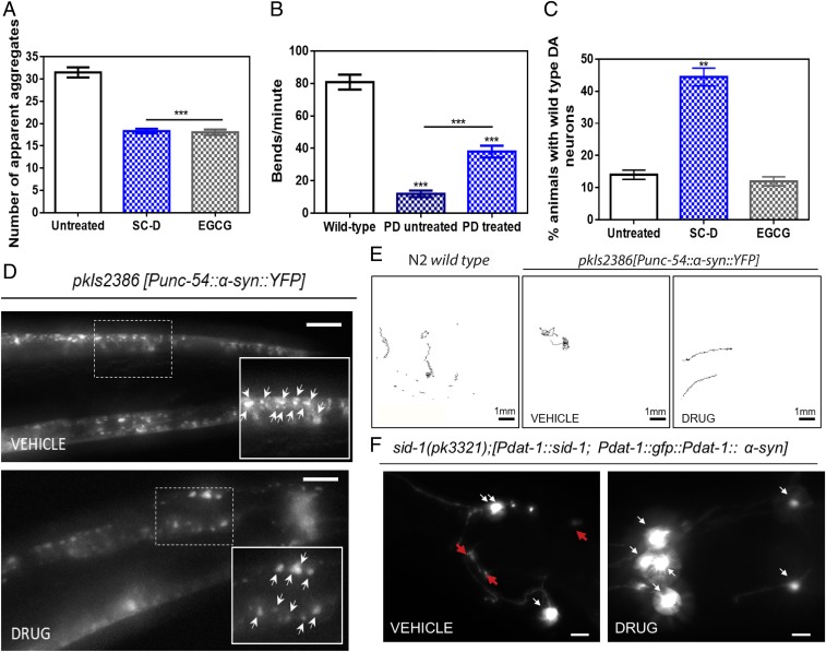
Fig. 5.
Inhibition effect of the compound in the formation of α-Syn inclusions and protection from the α-Syn–induced dopaminergic cell death in C. elegans models of PD. (A) Quantification of α-Syn muscle inclusions per area of NL5901 worms in the absence (white) and presence of SC-D (blue) or EGCG (gray). (B) Worm-thrashing representation as the number of bends per minute of N2 wild-type and NL5901 worms treated without (white) and with SC-D (blue). (C) Percentage of UA196 worms that maintain a complete set of dopaminergic neurons (four pairs located in the head) after treatment without (white) and with SC-D (blue) or EGCG (gray) for 7 d after the L4 stage. (D) Representative images of α-Syn muscle aggregates obtained by epifluorescence microscopy of NL5901 worms treated without (Top, vehicle) and with SC-D (Bottom, drug). (Scale bars, 10 μm.) Between 40 and 50 animals were analyzed per condition. Aggregates are indicated by white arrows. (E) Path representation of the mobility of N2 wild-type (Left, vehicle) and NL5901 worms grown without (Middle, vehicle) and with SC-D (Right, drug). (Scale bars, 1 mm.) (F) Representative worms expressing GFP–α-Syn specifically in DA neurons without (Left, vehicle) and with SC-D (Right, drug) for 7 d after L4. Healthy neurons are labeled with white arrows, whereas neurodegenerated or missing neurons are labeled with red arrows. (Scale bars, 30 μm.) Between 40 and 50 animals were analyzed per condition in each experiment. Data are shown as means, and error bars are shown as the SE of means; **P < 0.01 and ***P < 0.001.

