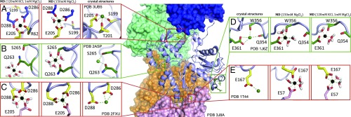
Fig. 2.
Review of divalent cation binding sites found in the PDB crystal structure library. F-actin is represented in the Middle (PDB 3J8A), in which four actin protomers are illustrated as surfaces (blue, green, orange, and pink), and the central protomer as a light blue cartoon. (A–E “crystal structures”) Enlargements of the cation binding sites from crystal structures are shown as green or yellow sticks, and Mg2+ and Ca2+ as black and green spheres, respectively. Sites that were previously described are boxed by red squares (A, C, and E) and those that were additionally observed in several crystal structures are boxed by green squares (B and D). (A–E “MD”) Mg2+ coordination after 100-ns MD simulation, in respective conditions. A and C are the upper and lower polymerization site, and E is the lower stiffness site (the upper site is identical and omitted for clarity).

