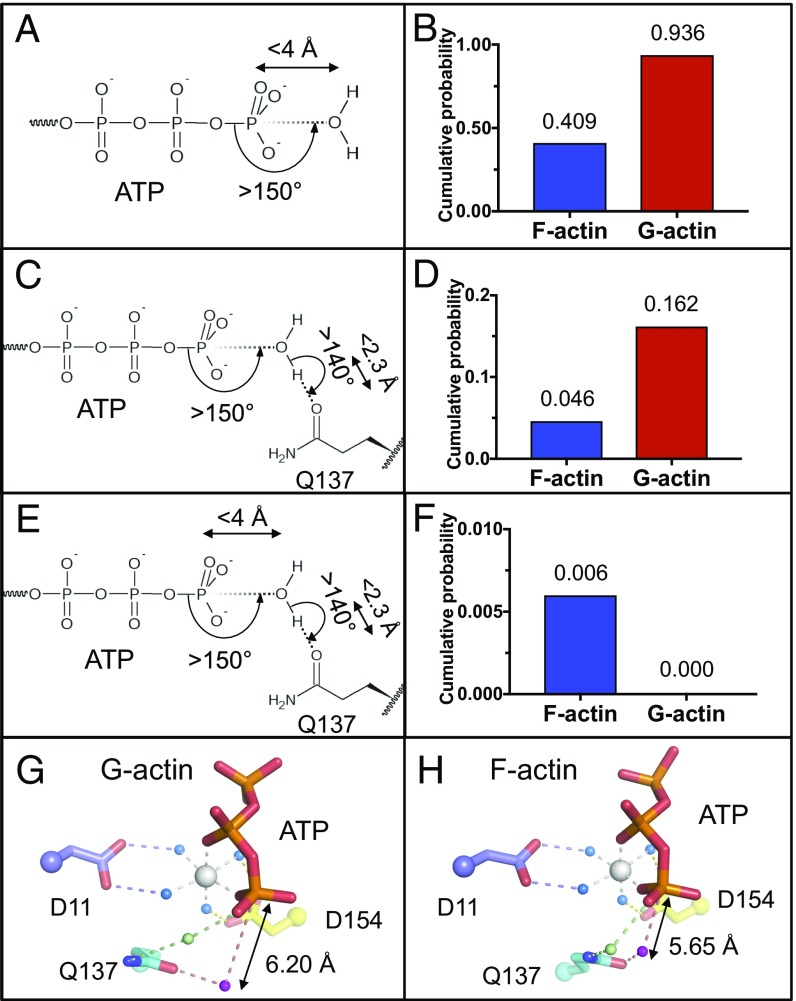
Fig. 4.
MD analysis of H2O close to the γ-phosphate and Gln137. (A, C, and E) Schematic representation of analysis conditions applied around ATP and Gln137. (B, D, and F) Cumulative probability of H2O with applied conditions from A, C, and E. (G) G-actin model, resulting from the combination of actin-DNaseI structure (PDB 1ATN) and ATP and ATP-bound Mg2+ of PDB 2V52. (H) Typical snapshot of MD simulation frame, satisfying all conditions from E. ATP and actin, sticks. Waters coordinated by Mg2+ are shown as blue, green, and purple spheres.

