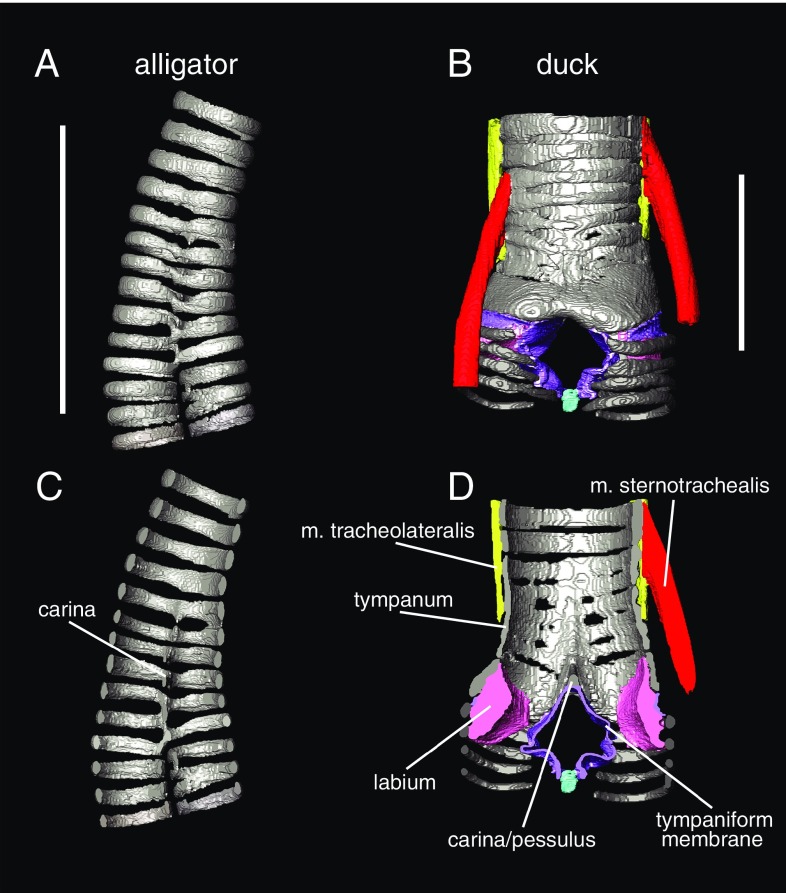
Fig. 2.
3D morphology of airway cartilage in archosaurs visible with diffusible iodine-based contrast-enhanced computed tomography (diceCT). 3D models of tracheal cartilage structure in the alligator (Alligator mississippiensis) (A and C) and Muscovy duck (Cairina moschata) (B and D) in gray. Panels show both external (A and B) and cross-sectional views of the tracheobronchial juncture (C and D). Soft tissue anatomy is clearly visible in B and D, including both intrinsic syringeal muscles (red and yellow), membranes (purple), and labia/vocal folds (pink). Specimens were dissected out, stained following ref. 109, and scanned at The University of Texas High-Resolution Computed Tomography Facility. Image segmentation was done in Avizo 6.3 (FEI Visualization Sciences Group). See ref. 8 for further details on staining and scanning parameters. (Scale bars: 2 cm.)

