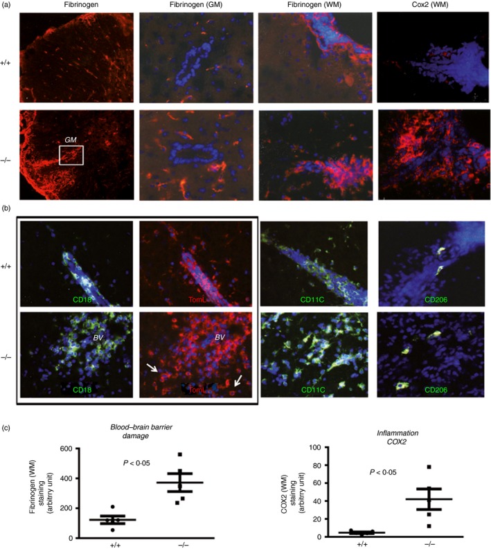
Figure 2.

Absence of CD93 exacerbates blood–brain barrier (BBB) damage and accumulation of polarized M1 macrophages (Cox2+, TL+, CD11c+ and CD206−) in experimental autoimmune encephalomyelitis (EAE). (a) Fibrinogen staining in the parenchyma (white or grey matters, WM/GM) was used as an indicator of BBB disruption and allowing transudation of other neurotoxic serum‐derived molecules (thrombin, complement). Robust Cox2 staining in perivascular inflammatory cells was found in CD93−/− EAE mice compared with discrete staining in wild‐type (WT) mice. (b) Double (CD18/TL) and single (CD11c or CD206) immunostaining was used to identify cytotoxic/pro‐inflammatory M1 versus regulatory/anti‐inflammatory M2 macrophage/microglia in EAE WT or CD93 mice. (c) Quantification of staining and image analysis were performed on five sections from three mice. Representative data obtained from tissue sections from three different brains from either WT or knockout (KO) animals with comparable clinical scores.
