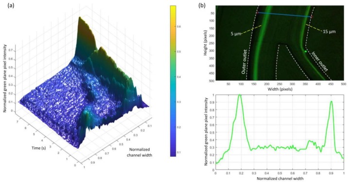
Figure 6.
(a) 3D green-plane intensity profile of experimentally focusing 5 μm particles when increasing flow rate from 0 to 5 mL·min−1 over a period of around 8 s; (b) 5 μm and 15 μm particles separation using the proposed trapezoidal spiral channel (top) image captured with the inverted fluorescence microscope at 5 mL·min−1 (bottom) green intensity profile shows the focusing peaks (each block on the x-axis is equal to 60 μm of the real channel width).

