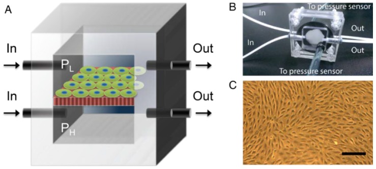
Figure 1.
Overview of the microfluidic platform. (A) Schematics of the device where podocytes were cultured on porous membrane, which was sandwiched by an upper chamber supplying PL and a lower chamber supplying with PH. The pressures in both chambers were controlled by peristaltic pumps that infused the medium via the inlet and outlet and monitored by pressure sensors. (B) An image of the real device; (C) a microscopic image of the differentiated podocytes monolayer. Scale bar, 50 μm.

