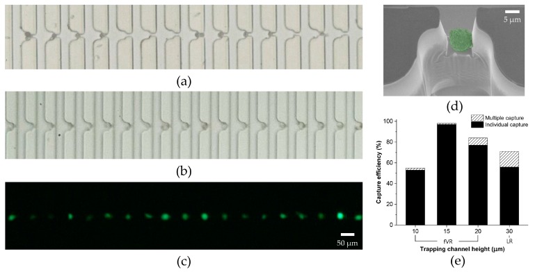
Figure 5.
Demonstration of the deterministic cell capture using LM2 MDA-MB-231: (a) bright field image of the tumor cell capture using an LR-type device; (b) bright field image and (c) fluorescent image of the individual tumor cell capture using an fVR-type device; (d) SEM image of a captured tumor cell from the fVR-type device; (e) capture efficiency of the LR- and fVR-type devices regarding various trapping channel heights. The graph also includes the individual capture rate.

