
Figure 3.
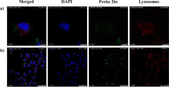
B16F10 cells were costained with 26c (4 μM) and LysoTracker Red and then analyzed under a confocal microscope. Nuclei were stained with DAPI. Blue: Nucleus; red: LysoTracker Red,; green: 26c. Micron bar: (a) 10 μm; (b) 50 μm.
Official websites use .gov
A
.gov website belongs to an official
government organization in the United States.
Secure .gov websites use HTTPS
A lock (
) or https:// means you've safely
connected to the .gov website. Share sensitive
information only on official, secure websites.
B16F10 cells were costained with 26c (4 μM) and LysoTracker Red and then analyzed under a confocal microscope. Nuclei were stained with DAPI. Blue: Nucleus; red: LysoTracker Red,; green: 26c. Micron bar: (a) 10 μm; (b) 50 μm.