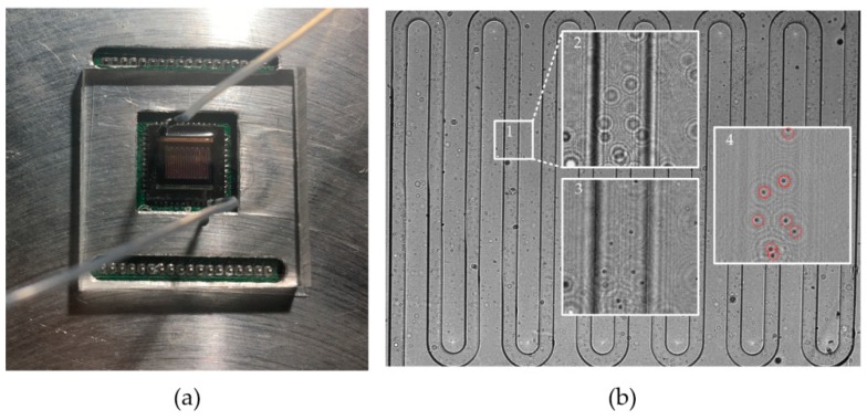
Figure 2.
The proposed flow cytometer: (a) The flow cytometer system; (b) the holograms of the microfluidic chip captured by the system. Box 2 is an amplificatory image of box 1, and box 3 is a diffractive reconstruction image of box 2. Box 4 is the image of box 3 with the background removed, where the red circles mark the cells.

