
Figure 6.
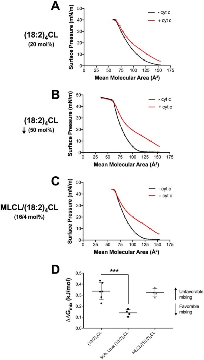
Proteolipid domain organization in biomimetic membranes is disrupted with the loss of cardiolipin concentration due to favorable Gibbs-free energy of mixing. Biomimetic monolayers were constructed containing (A) 20 mol % (18:2)4CL, (B) 50 mol % reduction of (18:2)4CL, or (C) 16/4 mol % MLCL/(18:2)4CL. Biomimetic monolayers were composed of (18:0–22:6)PC/(16:0–20:4)PE/CL/DOPI/DOPS/Chol in the absence or presence of 3.8 μm cytochrome c. The Gibbs-free energy of mixing (D) was calculated at a physiologically relevant surface pressure of 30 mN/m, in the absence and presence of cyt c. The ΔΔG (i.e. ΔG(+)protein − ΔG(−)protein) of lipid mixing (D) is presented where a higher, more positive, ΔΔGmix value indicates unfavorable lipid-lipid mixing. Data are average ± S.D. from four to six independent experiments. Asterisks indicate significance from monolayers containing (18:2)4CL: ***, p < 0.001.