Figure 5.
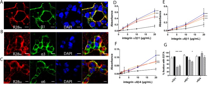
R28Nt and its subdomains interact with integrins α3β1, α6β1, and α6β4. A–C, immunostaining of R28Nt and different integrin monomers on HEC-1-A cells: β1 (A), α3 (B), and α6 (C). Red, R28Nt; green, the specified integrin monomer; blue, DAPI; far-right image, merge. Scale bars correspond to 5 μm. D–F, assessment of integrin binding: α3β1 (D), α6β1 (E), and α6β4 (F) binding to R28Nt (red), R28-N1 (blue), R28-N2 (orange), or BSA (black) by ELISA. Error bars correspond to S.E. of three to five independent experiments (two-way ANOVA at 20 μg/ml with Bonferroni post-tests against BSA). G, role of divalent cations in the binding of integrins to R28Nt, PBS (white column); PBS + EDTA (black column); PBS + Mn2+ (gray column); and PBS + Ca2+ (hatched column) (one-way ANOVA of four independent experiments). *, p < 0.05; ***, p < 0.001.

