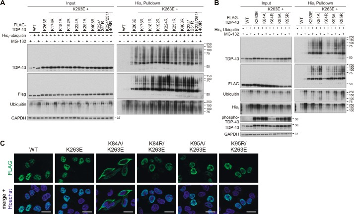
Figure 6.
Investigation of ubiquitinylation sites for the hyper-ubiquitinylated mutant TDP-43K263E. HEK293E cells were transfected with FLAG–TDP-43WT and FLAG–TDP-43K263E alone or combined with the indicated lysine substitutions localized in the C-terminal part (A) or the NLS sequence (B). His6-ubiquitin (+) or vector control (−) was cotransfected, as indicated. Cells were then treated with MG-132 (+) or DMSO (−) for 2 h. The urea-soluble lysates were prepared, and His6-ubiquitin–conjugated proteins were pulled down from cell lysates. Total cell lysates (Input) and Ni-NTA–agarose eluates were analyzed with Western blotting and stained for FLAG, TDP-43, ubiquitin, and GAPDH, and in B additionally with anti-His6. Lysates samples in B were re-run and subjected to Western blot analysis of phospho-TDP-43. C, HEK293E cells overexpressing FLAG–TDP-43WT and FLAG–TDP-43K263E alone or combined with the indicated Lys-84 and Lys-94 substitutions were immunostained with rabbit anti-FLAG (green). Nuclei were counterstained with Hoechst 33342 (blue). Scale bars correspond to 20 μm.

